
Research use only. Not for clinical use.
Body Composition
Skeletal muscle, subcutaneous, visceral and intermuscular fat on CT and MRI, as cross-sectional areas and volumes.
CT Dice against board-certified radiologists
- Skeletal muscle
- 96.9%
- Subcutaneous fat
- 97.0%
- Visceral fat
- 96.7%
- Intermuscular fat
- 89.2%
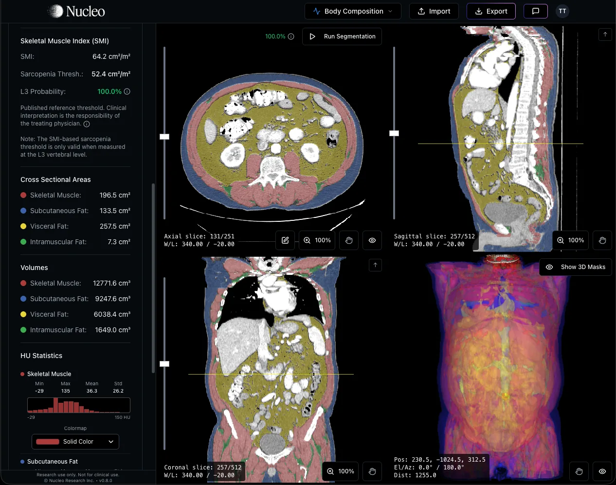
Four compartments, measured
Finds L3 for you
Fast
Why body composition
Body composition, the balance of skeletal muscle and visceral, subcutaneous and ectopic fat, is associated with clinically meaningful outcomes across many settings. Low skeletal muscle mass has been linked to chemotherapy tolerance, peri-operative risk, and survival in oncology, and to frailty and mortality in older adults. Visceral and ectopic fat have been linked to cardiovascular and metabolic risk, including type 2 diabetes and fatty liver disease. With the growing use of weight-loss therapies such as GLP-1 agonists, distinguishing fat loss from muscle loss has also become clinically relevant. This information is already contained in the CT and MRI scans acquired during routine care, yet it is rarely quantified in practice: extracting it requires manually identifying the correct level and delineating each tissue compartment, a slow, manual step that does not fit routine reporting volumes.
Cross-sectional areas at the third lumbar vertebra (L3) are the most widely used single-slice measure (Mourtzakis et al., 2008), CT and MRI tissue areas have been validated against cadaver dissection (Mitsiopoulos et al., 1998), and low skeletal muscle on CT predicts survival in cancer independently of body mass index (Prado et al., 2008; Martin et al., 2013). Segmenting by hand is slow and varies between readers, which limits its use in large studies (Paris et al., 2020).
1.44 times the risk of death
Across 38 studies and 7,843 patients with solid tumours, low skeletal muscle on CT was linked to a 44% higher risk of death.
38 of 250 obese patients
Among obese patients with lung or gastrointestinal cancer, 15% had sarcopenia that their weight concealed. It independently predicted death (hazard ratio 4.2).
0.92 c statistic
In 1,473 patients, a survival model using body composition on CT and weight history predicted survival better than stage, diagnosis, age and performance status.
Four tissues, measured on every CT slice
Nucleo finds L3 automatically, with an L3 probability shown for every slice, and outlines the four tissues.
At L3 you get the area and density of each tissue, and skeletal muscle index once the patient’s height is entered. Across the scan you get the volume of each tissue. Published sarcopenia thresholds are shown for reference; the software does not classify sarcopenia.
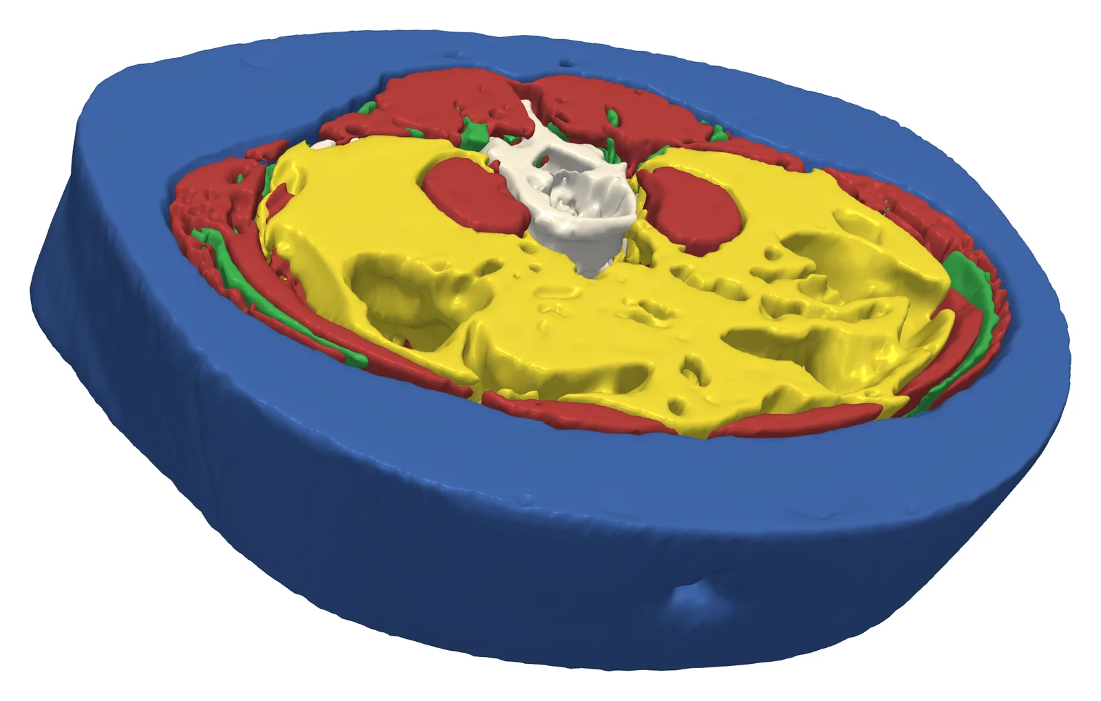
- Skeletal muscle
- Subcutaneous fat
- Visceral fat
- Intermuscular fat
Correct anything, keep the record
Brush, eraser, polygon and smart fill tools work directly on the masks. The brush only paints tissue in the HU range of the compartment you are editing, the measurements update as you go, and every export keeps the original values beside the edited ones.
Accuracy and speed
- 96.9%1
- Skeletal muscle Dice
- 97.0%1
- Subcutaneous fat Dice
- 96.7%1
- Visceral fat Dice
- 89.2%1
- Intermuscular fat Dice
- >2,500x2
- Faster than manual segmentation
- 0.2 s3
- Per slice
- DSC against board-certified radiologists on 226 CT subjects. On MRI: 82.5% muscle, 80.5% subcutaneous fat, 76.2% torso fat.
- Estimated 10 minutes per manual segmentation.
- CT analysis run in Chrome on an Apple M1 Max laptop. Single-slice analysis is not available for MRI.
Fits your workflow
In the browser
Scans never leave your computer.
On your hospital network
Patient data stays in the hospital.
Questions
Do scans leave my computer?
Not by default. Analysis runs in the browser or the desktop app. Cloud processing is an option you can turn on for large series.
Which CT scans does it work on?
Routine abdominal and whole-body CT. It was validated on contrast-enhanced portal venous and non-contrast low-dose CT covering the whole body, the abdomen and pelvis, or the abdomen.
Can I change the slice or the outlines?
Yes. You can run the analysis on any slice and correct any mask with the editing tools. Exports keep the original values beside the edited ones.
See it on your own scans.
We can walk through Body Composition on a real study and show the outputs end to end.
Book a demo